Full Skin Examination

What is a Full Skin Examination Used to Assess?
A full skin examination is performed on patients to assess pigmented and non pigmented skin.
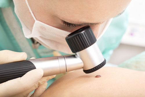
This full skin check ( from head to toe ) is undertaken with a hand-held Dermoscope.
Dermoscopy refers to the examination of the skin with a hand-held device using surface microscopy. It allows the doctor to visualize lesions in much greater clarity, and making diagnoses based on pattern and pigment recognition.
It has reduced the need greatly for skin biopsy and excision.
It is used in the evaluation of pigmented and non-pigmented skin lesions.
In experienced hands, it will differentiate between benign moles, atypical moles and malignant ones ( melanoma ).
It very useful in the diagnosis of pre-malignant skin cancers and non-melanoma-skin-cancers.
It is also a very useful took in the diagnoses of common rashes and inflammatory and infectious skin conditions.
Following a complete check, patients are quite often reassured and advised on skincare .
If abnormalities are detected, follow-up biopsies, excisions or tertiary referrals are made.
Depending on clinical risk, appointment intervals are estimated and follow-up appointments made.
What are the risk Factors for malignant melanoma and skin cancer?
Risk factors for malignant melanoma and skin cancer include the following:
- Fair complexion
- History of blistering sun burn in the past
- Positive family history of malignant melanoma in a first degree relative
- Immunosuppressive illness
- Sun bed use
- Occupational risk
- Previous skin cancer
